
Figure 3.
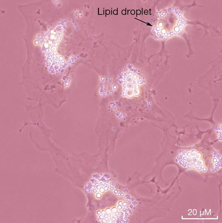
Light micrograph of an overnight culture of primary HSCs obtained from 3 month-old wild type C57BL/6J mice fed a retinoid-sufficient chow diet throughout its entire life. The HSCs were isolated employing standard procedures involving pronase E perfusion of the liver followed by floatation of HSCs through a Nycodenz gradient. Freshly isolated HSCs taken from the Nycodenz gradient were cultured on plastic dishes and maintained overnight in Delbecco’s modified Eagles medium containing 10% (v/v) fetal bovine serum. The numerous cytoplasmic lipid droplets are the site of retinyl ester storage within these cells and a distinguishing characteristic of HSCs. Similar lipid droplets present in PSCs are a site of retinyl ester storage in the pancreas. If culture of these cells on the plastic dishes were continued for one week or more, the HSCs will activate and the retinoid-containing lipid droplets will be totally lost. HSCs, hepatic stellate cells.