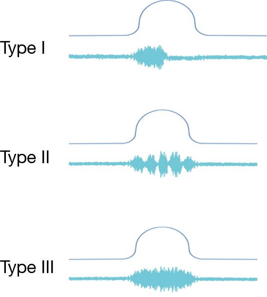
Figure 2.

Three types of DSD. The top line shows a Pdet tracing during an attempted void while the bottom line shows EMG activity. Type I shows a temporary failure of the EMG to silence during contraction while type II is intermittent but not obstructing. Type III shows a crescendo of the EMG throughout the detrusor function. DSD, detrusor sphincter dyssynergia; Pdet, detrusor pressure; EMG, electromyography.
