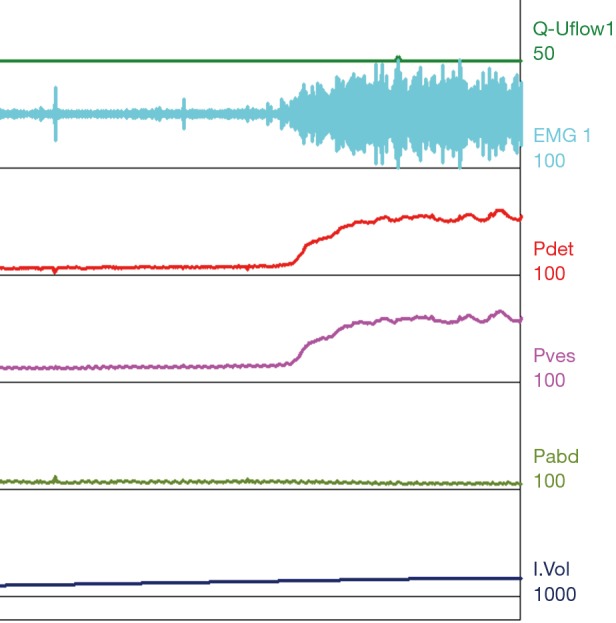
Figure 7.

Type III DSD. Patient with normal compliance and a decreased capacity of 160 mL. At time of void initiation the EMG shows a crescendo, decrescendo firing pattern throughout the void, elevation of Pdet and with no appreciable voided volume. DSD, detrusor sphincter dyssynergia; EMG, electromyography; Pdet, detrusor pressure; Pves, intravesical pressure; Pabd, abdominal pressure.
