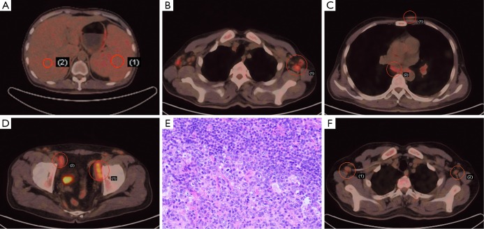
Figure 1.
18F-FDG PET/CT and histology findings of MCD in a 43-year-old male. (A) 18F-FDG PET/CT demonstrated slight FDG metabolism of spleen and liver. Glucose uptake is little higher in spleen (SUVmax =2.3, right) than that of liver (SUVmax =2.0, left); (B) 18F-FDG PET/CT showed increased FDG metabolism of lymph nodes in axilla (SUVmax =3.9); (C) 18F-FDG PET/CT revealed subcutaneous nodules and lymph nodes in mediastinum with higher glucose uptake (SUVmax =1.8, upper; SUVmax =3.0, lower); (D) enlarged lymph nodes on both sides of the pelvic wall on 18F-FDG PET/CT (SUVmax =5.2, right; SUVmax =3.7, left); (E) the histological results of the right inguinal lymph nodule biopsy (HE, ×200); (F) following 3 cycles of R-CHOP regimen, 18F-FDG PET/CT showed shrunken lymph nodes in both axillas and decreased FDG metabolism (SUVmax =2.5).

