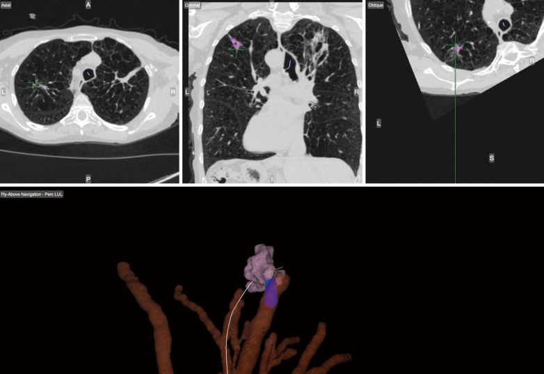
Figure 4.
CT with multiple views (top) and computer software generated map (bottom) of airway anatomy with guidance (white line) to the target lesion in the right upper lobe (purple). The blue cylinder represents the trackable biopsy instrument used for parenchymal sampling under navigation guidance.

