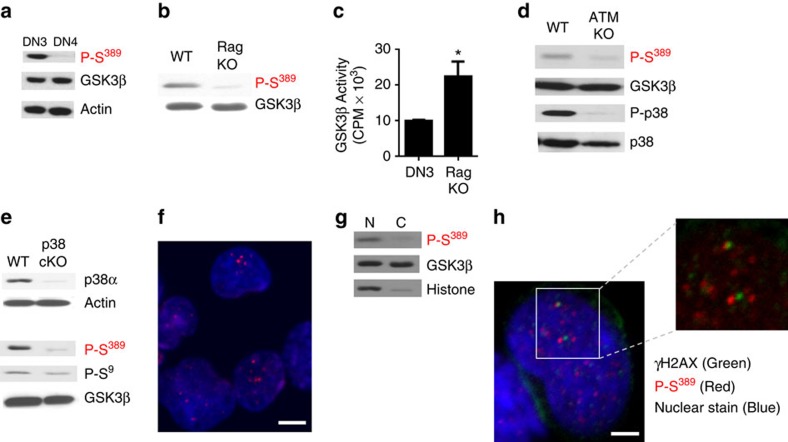
Figure 2. DSBs generated by V(D)J in developing T cells induce inactivation of GSK3β by p38 MAPK.
(a) DN3 and DN4 thymocytes were examined for P-S389 GSK3β and total GSK3β by western blot analysis. Actin is shown as a loading control. (b) The levels of P-S389 GSK3β and total GSK3β in DN3 thymocytes from WT and Rag KO were examined by western blot analysis. (c) GSK3β activity in lysates from DN3 thymocytes from WT and Rag KO mice were determined using in vitro kinase activity assays (n=3,±s.e.m.). *P<0.05 as determined by t-test. (d) The levels of P-p38 MAPK, total p38 MAPK, P-S389 GSK3β and total GSK3β in DN3 thymocytes from WT and ATM KO mice were determined by western blot analysis. (e) DN3 thymocytes from WT and p38α conditional KO (p38cKO) mice were examined for p38α, P-S389 GSK3β, P-S9 GSK3β and total GSK3β by western blot analysis. Actin is shown as a loading control. (f) DN3 thymocytes were examined by immunostaining and confocal microscopy for the presence of P-S389 GSK3β (red) and TOPRO nuclear stain (blue). Scale bar, 5 μm. (g) Western blot analysis for P-S389 GSK3β and total GSK3β using nuclear and cytosolic extracts from DN thymocytes. Histone is shown as a marker for the nuclear fraction. (h) DN3 thymocytes were examined by immunostaining and confocal microscopy for the presence of γH2AX (green), P-S389 GSK3β (red) and TOPRO nuclear stain (blue). Scale bar, 2 μm. Data are representative of three or more independent experiments.

