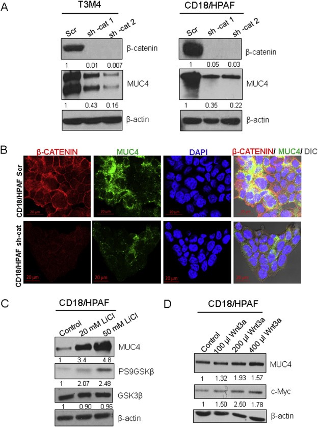
Figure 2.

MUC4 protein and RNA expression are governed by β‐catenin. (A) CD18/HPAF and T3M4 pancreatic ductal adenocarcinoma (PDAC) cell lines were transfected with two lentiviral shRNAs that targeted β‐catenin (shRNA‐cat1, shRNA‐cat2) or a scrambled sequence in the PLKO.1 vector. Further, knock down of β‐catenin resulted in reduced MUC4 protein expression. (B) Confocal microscopy analysis was used to analyze MUC4 (green) and β‐catenin (red) levels in CD18/HPAF Scr and CD18/HPAF shRNA‐β‐catenin (shRNA‐cat) cells. (C) Treatment with lithium chloride (LiCl, a GSK3‐β inhibitor) at 20 mM and 50 mM concentrations was used to induce nuclear β‐catenin. Western blot analysis showed a dose‐dependent effect on MUC4 levels and an increase in phosphorylation of the inhibitory Ser9 residue of GSK3‐β, while total GSK3‐β levels were unaffected. (D) CD18/HPAF cells were treated with increasing amounts of Wnt‐3A‐conditioned medium; levels of c‐Myc were used as a positive control.
