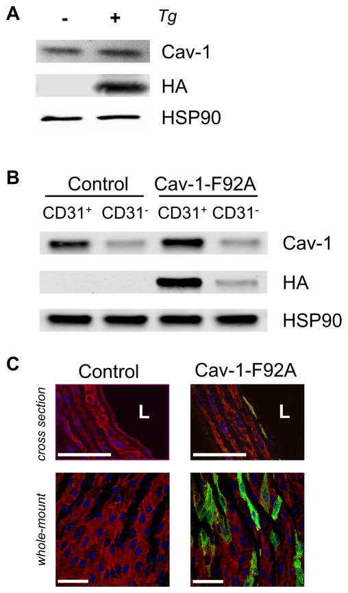
Figure 1. Expression of the Cav-1-F92A-HA transgene.
A, Whole lung protein from control and Cav-1-F92A mice was analyzed for the expression of the transgene. B, MLECs were isolated with CD31-dynabeads. The fraction bound (CD31+) and the non-bound fraction (CD31−) was separated and immunoblotted for HA. The endothelial specific protein VECAD was used as a marker for the enrichment of endothelial cells by using the CD31-beads. C, Top panel, Cross sections of the thoracic aorta. Sections were stained for the nucleus (blue), α-SMA (red) and HA (green) [L, lumen]. Bottom panel, Whole-mount staining of mesenteric artery. En-face preparations were stained for the nucleus (blue), PECAM-1 (red) and HA (green). Scale bars are 50 μm.

