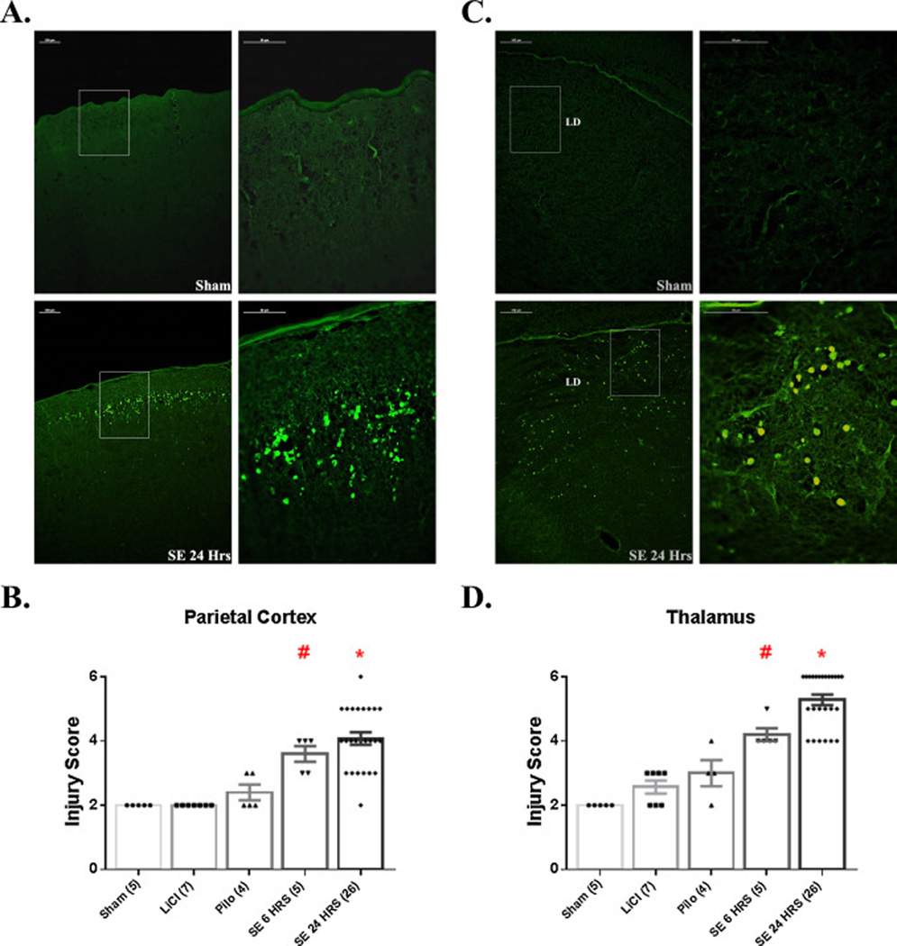
Figure 4.
SE induces neuronal damage in neocortex and thalamus. (A) Images of FJB staining in parietal cortex of Sham and SE24Hrs P7 pups. The image on the right of each row is a higher magnification of the boxed area on the image on the left. As shown, SE induces significant neuronal injury predominantly in layer 2 of the cortex. (B) Graph depicting extent of neuronal injury in parietal cortex, based on a 6-point scoring system. # p<0.05: SE 6 HRS vs. Sham, LiCl, Pilo (analyzed by Mann-Whitney). * p<0.05: SE 24 HRS vs. Sham, LiCl, Pilo (Mann-Whitney test). (C) Images of FJB staining in thalamus (lateral dorsal nucleus) of Sham and SE24Hrs P7 pups. The image on the right of each row is a higher magnification of the boxed area on the image on the left. SE results significant neuronal injury at 6 and 24 h post-SE. # p<0.05: SE 6 HRS vs. Sham, LiCl, Pilo (Mann-Whitney test). * p<0.05: SE 24 HRS vs. Sham, LiCl, Pilo, SE 6 HRS (Mann-Whitney test). Abbreviations: LD=lateral dorsal thalamic nucleus. Scale bars: (A, C) left column=100 µm (low magnification images) and right column 20 µm (high magnification images).

