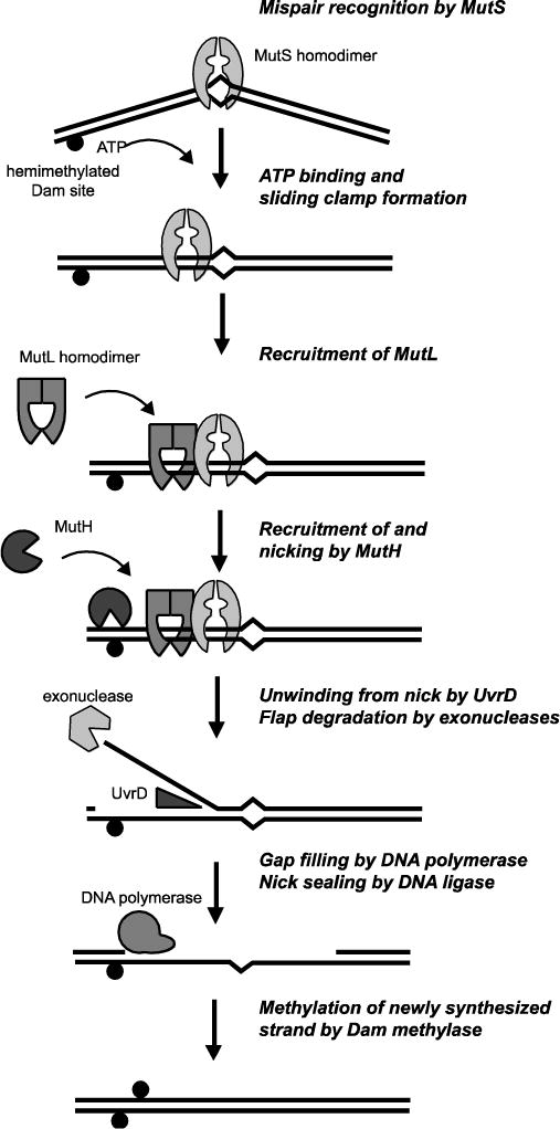
Fig 1. Diagram of methyl-directed MMR.

Main steps in the E. coli methyl-directed MMR pathway (see main text). Black circle indicates the presence of a methylated adenosine at a d(GATC) site.

Main steps in the E. coli methyl-directed MMR pathway (see main text). Black circle indicates the presence of a methylated adenosine at a d(GATC) site.