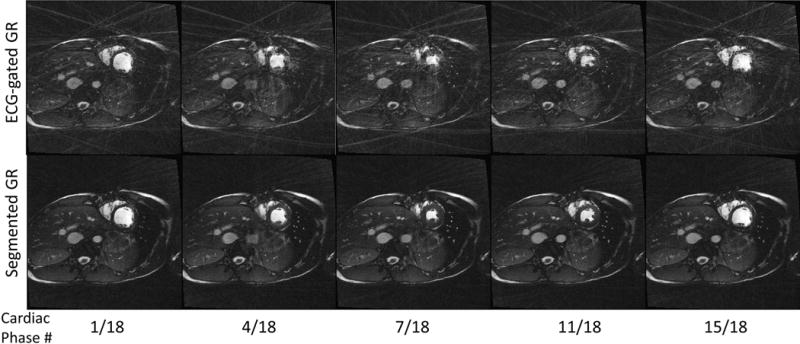
Figure 5.

Comparison between 2D radial ECG-gated cardiac cine images using the proposed segmented GR (bottom row) and conventional GR (top row). Five out of 18 cardiac phases are chosen for display. Severe streaking artifacts due to the non-uniform sampling of the k-space found in conventional GR are absent in the reconstructed images using the proposed segmented GR method.
