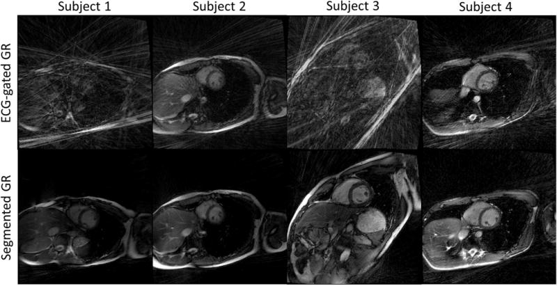
Figure 7.

3D cardiac CINE short axis images of four healthy volunteers. The images generated from conventional GR (subject 1 and 3) are non-diagnostic due to severe streaking artifacts. The images using segmented GR method provide uniformly good quality even though the same number of radial spokes were used.
