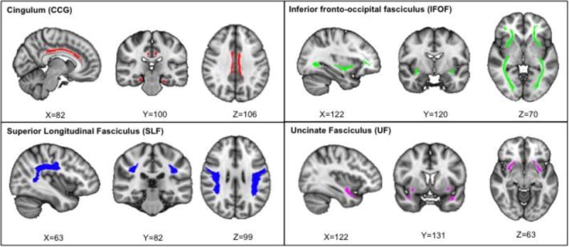
Figure 1.

AD-related white matter tracts: the cingulum (red, top left), the inferior fronto-occipital fasciculus (green, top right), the superior longitudinal fasciculus (blue, bottom blue), and the uncinate fasciculus (pink, bottom right). Tract representation is shown using the standard MNI brain in radiological orientation.
