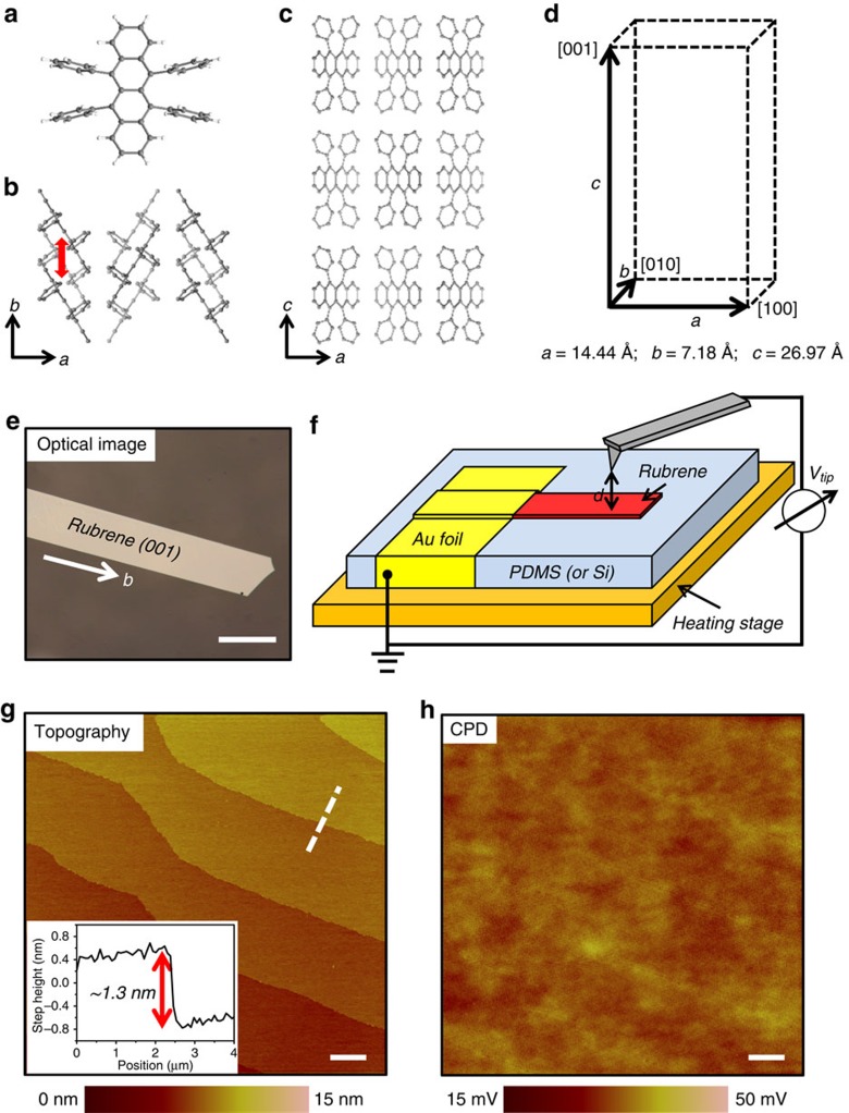
Figure 1. Crystal structure and SKPM measurement of rubrene single crystals.
(a) Molecular structure of rubrene. (b) Crystal structure in the a-b plane; red arrow indicates the π-stacking interaction. (c) Crystal structure in the a-c plane. (d) Orthorhombic structure and lattice parameters of rubrene. (e) Optical micrograph of as-grown rubrene crystal. Length of scale bar, 200 μm. (f) SKPM setup for CPD measurement. The sample sits on top of a heating stage and is grounded through gold foil. The conducting probe scans in a two-pass ‘lift mode' and a constant lift height (d=50 nm) is used in the ‘interleave' pass. (g) Topography of rubrene single crystal shows typical terrace structure and each terrace has height corresponding to one molecular layer. Length of scale bar, 2 μm. Inset: step height profile of the dashed line. (h) CPD image obtained simultaneously with topography shows nearly homogeneous CPD across the surface. Length of scale bar is 2 μm.

