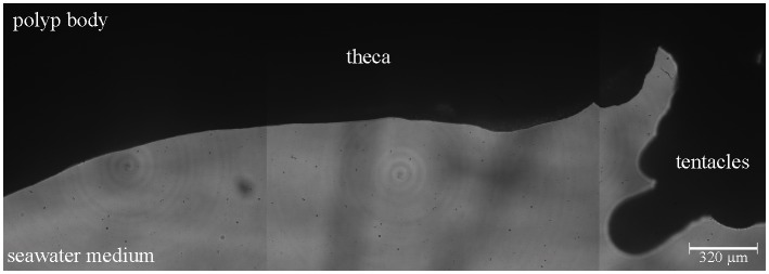
Fig 2. Example of a coral polyp not exposed to stimuli.
A merged hologram image is shown for one polyp along the observation path. Dark areas = polyp (opaque to the DHM LED beam), light area = surrounding seawater medium (transparent to the DHM LED beam). No mucus layer or strings were detected along any part of the observation path.

