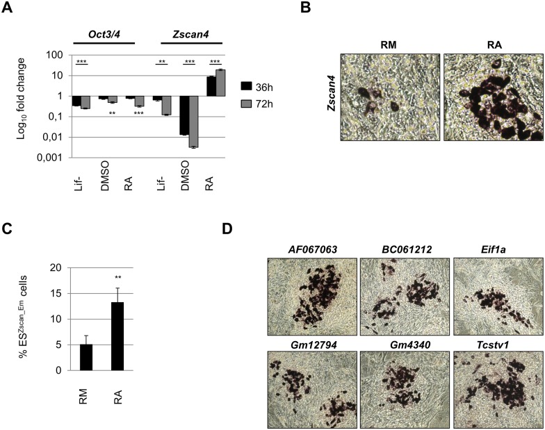
Fig 1. RA induces Zscan4 ESC metastate.
(A) ESCs were cultured for 36 and 72 h in differentiating conditions: Lif-, 1% DMSO and 1.5 μM RA. The mRNA expression levels were assessed by qRT-PCR and normalized to RM condition. The average and SD of duplicate samples from each of three independent biological replicates are shown: **, p < .01; ***, p < .001, in a Student’s t test. (B) Zscan4 expression pattern in ESCs cultures by RNA in situ hybridization upon 5 days of treatment in RM or RA. RNA ISH showed “spotted” patterns on ESC colonies (40x). (C) Percentage of RM-Zscan4+ and RA-Zscan4+ cells was evaluated by flow cytometry analyses. The mean % ESZscan4_Em cells ± SD of three independent experiments is presented with statistical analysis performed using Student’s t test (**, p < .01). (D) Distribution of Zscan4 related gene signature in ESC cultures by RNA in Situ hybridization showed “spotted” patterns on ESC colonies. Most representative colonies were magnified (20x) to show the detailed staining patterns.

