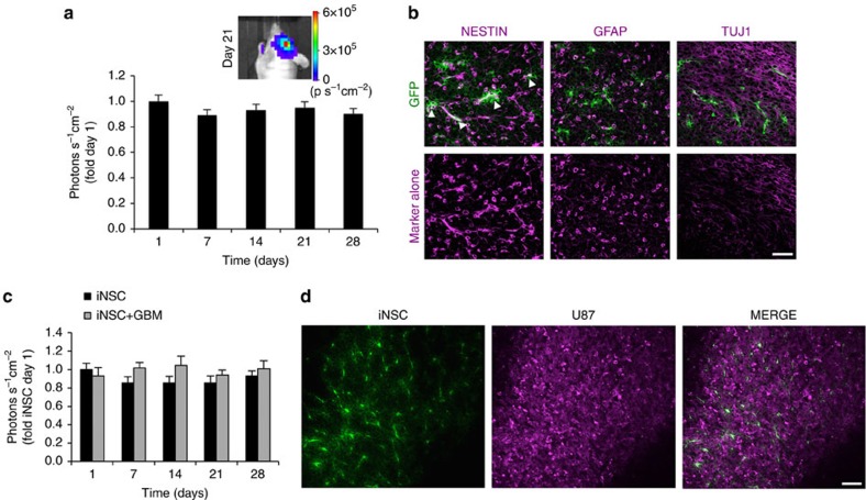
Figure 2. In vivocharacterization of iNSCs transplanted in the mouse brain in the presence or absence of GBM.
iNSC-GFPFLs were implanted into the frontal lobe of mice in the presence or absence of U87 human GBM (n=6). Serial bioluminescence imaging was used to monitor the volumes of iNSC-GFPFL. Two weeks after implantation, a subset of mice was killed and their brains sectioned and analysed. (a) Summary graph depicting the volume of iNSC-GFPFL in the brain in the absence of GBM through 28 days. (b) Immunofluorescence analysis of iNSC-GFPFL (green) 14 days post implantation into the brain. Nestin+ iNSC-GFPFLs were detected (indicated by arrowheads), and iNSC-GFPFL differentiation was detected with GFAP or Tuj-1 staining shown in magenta. Representative fluorescent images showing only the red (555 nm) secondary antibody channel are shown in the bottom row. (c) Engraftment of iNSC-GFPFL cells implanted in the brain in the presence or absence of human GBM (n=10 per group) measured using bioluminescence imaging. (d) Representative fluorescence imaging of post-mortem tissue sections showing GFP+ iNSCs (green) are still present in mCherry+ GBMs (magenta) 28 days after implantation. Scale bars in b,d, 40 and 50 μm, respectively. Data are mean±s.e.m. In a, P>0.05 by repeated measures one-way ANOVA. In c, P>0.05 by repeated measures two-way ANOVA.

