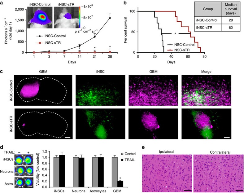
Figure 6. Antitumour efficacy of iNSC-sTR treatment of orthotopic human GBM xenografts.
Human U87 GBM cells expressing mCherry and firefly luciferase were implanted into the parenchyma of mice with iNSC-sTR or control iNSC-GFP. (a) Serial bioluminescence imaging showing the growth of the human GBM xenografts treated with iNSC-sTR or control iNSC-GFP through 28 days (n=12 per group). (b) Kaplan–Meier curves showing the survival of U87-bearing animals treated with iNSC-sTR or control iNSC (n=12 per group). (c) Representative fluorescent micrographs of brain sections from control- or iNSC-sTR-treated mice bearing mC-FL-expressing U87 tumours. Magenta=U87; Green=iNSC-sTR or iNSC-GFP. (d) Images and summary data showing the viability of iNSCs, neurons, astrocytes and U87 GBM cells cultured with conditioned media from iNSC-sTR or iNSC-GFP. (e) Representative histological images of the brain sections from mice injected intracranially with iNSC-sTR (n=5). Images were captured 0.5 mm from the implantation site (ipsilateral) and the same distance from the midline in the opposite hemisphere (contralateral). Scale bar in c, 1,000 and 30 μm in magnification. Scale bar e, 30 μm. Data are mean±s.e.m. Data in a,b,d are from three independent experiments. Data in a,b are n=12 per group. In a, *P<0.05 by repeated measures two-way ANOVA. In b, *P<0.01 by exact log-rank test. In d, *P<0.01 by Student's t-test.

