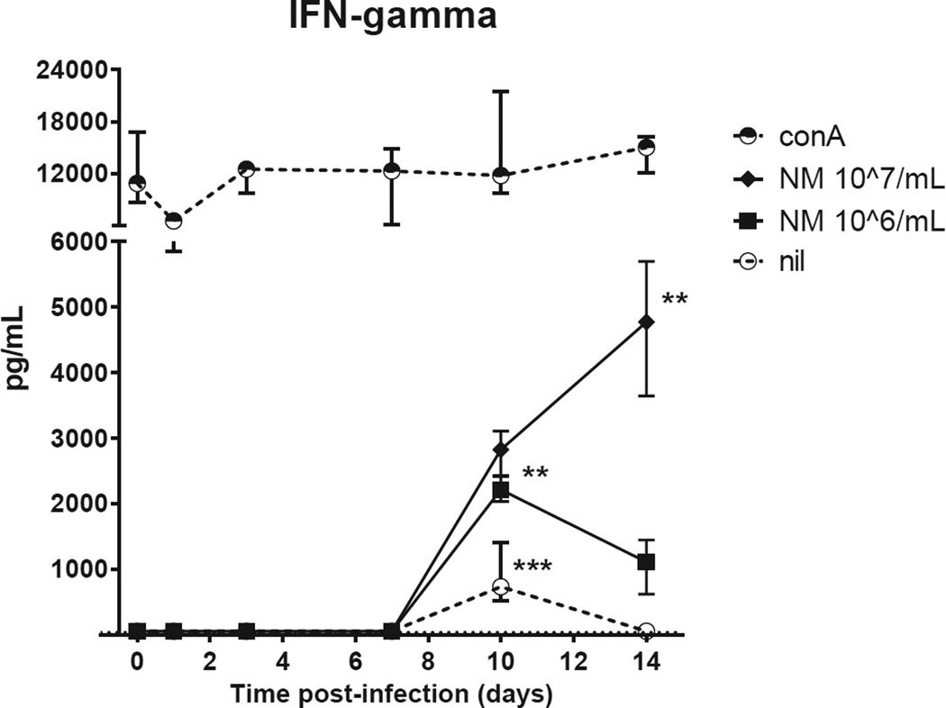
Figure 4. Early IFN-γ production by stimulated splenocytes of C. burnetii-infected immunocompetent BALB/c mice.
Splenocytes were stimulated for 48h with either conA [2.5 µg/mL], NM phase I [10^7/mL], NM phase I [10^6/mL], or left unstimulated (nil). The median ± IQR cytokine production is shown per time point of four pairs of infected mice. T=0 shows the median ± IQR of five pairs of uninfected mice. P values were calculated by Kruskal-Wallis test followed by Dunn’s multiple comparison test comparing cytokine concentrations of infected mice at different time points with uninfected mice. * P<0.05, ** P<0.01, *** P<0.001.
Abbreviations: conA, concanavalin A; NMI, C. burnetii Nine Mile phase I.

