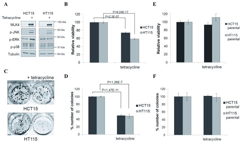
Figure 4.
Reintroduction of MLK4-WT decreases cell viability. A, Expression of MLK4-WT in HCT15 and HT115 was induced by tetracycline for 1 day. WCL were analyzed by western blot. B, Expression of MLK4-WT in HCT15 and HT115 was induced by tetracycline for 4 days. Viability was determined by MTT assay. Error bars indicate ±SEM from three independent experiments performed in triplicate (n=9). C and D, HCT15 and HT115 were seeded at low density and expression of MLK4 was induced by tetracycline. After 2 weeks cells were stained with crystal violet (C) and results were quantified by absorbance (D). Error bars indicate ±SEM from three independent experiments (n=3). E, Parental HCT15 and HT115 were treated with tetracycline for 4 days. Viability was determined by MTT assay. Error bars indicate ±SEM from three independent experiments performed in triplicate (n=9). F, Parental HCT15 and HT115 were seeded at low density and expression of MLK4 was induced by tetracycline. After 2 weeks cells were stained with crystal violet and results were quantified by absorbance. Error bars indicate ±SEM from three independent experiments (n=3).

