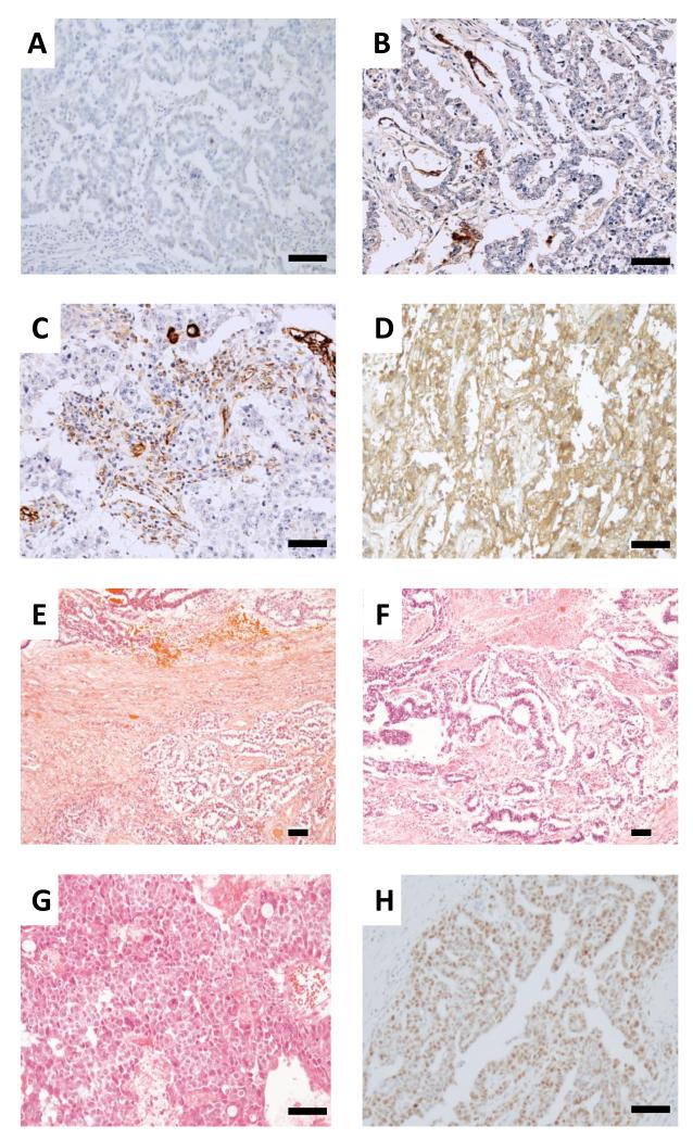
Figure 1. Representative staining of stage I non-seminomatous germ cell tumor samples.
Immunohistochemistry for CXCL12 staining A negative B <10% C ~30% and D 100% positive. E Haematoxylin and Eosin staining showing a combined seminomatous (bottom of photomicrograph) and non-seminomatous tumour (top) composed of less than 10% embryonal carcinoma. F Tumor composed of 25% embryonal carcinoma and 75% yolk sac tumor. The yolk sac and embryonal carcinoma are intermingled in a polyembryo matous fashion, mimicking the earliest stages of embryonic development. G Tumor entirely composed of embryonal carcinoma. H Embryonal carcinoma showing 75% positivity for MIB1 immunohistochemistry. (Scale bar, 100 microns).

