Fig. 9.

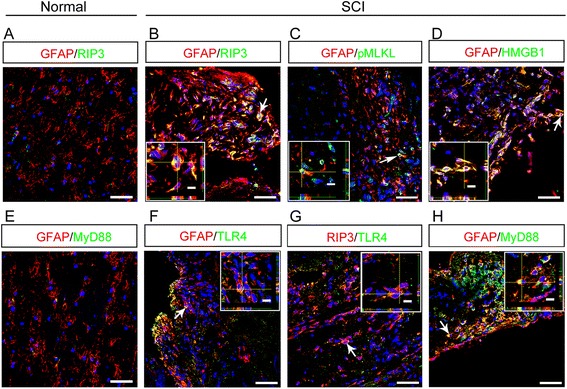
Expression of necroptotic markers and TLR/MyD88 by astrocytes in injured human spinal cord. a Double-staining of GFAP with RIP3 in uninjured human spinal cord. Notice the very weak expression of RIP3. Bar = 50 μm. b–d Representative images of double-staining of GFAP with RIP3, pMLKL and HMGB1 in human spinal cord at 5 days post-injury. Notice the co-localization of RIP3, pMLKL and HMGB1 with GFAP. Bars = 50 μm. e Double-staining of GFAP with MyD88 in uninjured human spinal cord. Notice the very weak expression of MyD88. Bar = 50 μm. f–h Representative images of double-staining of TLR4/RIP3, TLR4/GFAP and GFAP/MyD88 in human spinal cord at 5 and 15 days post-injury. Notice the co-localization of TLR4 with RIP3 and GFAP, and MyD88 with GFAP. Bars = 50 μm
