
Fig. 3.
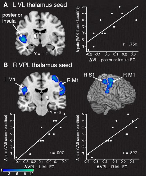
Correlations between changes in FC and changes in clinical pain after sham transcranial direct current stimulation (tDCS). a Decreased FC between the left VL thalamus (seed in white) and left posterior insula was correlated with a reduction in clinical pain after sham tDCS. b Decreased FC between the right VPL thalamus (seed in white) and left M1, right M1, and right S1 correlated with reduced clinical pain after sham tDCS. Data shown are Fisher-transformed r values. VL ventral lateral, VPL ventral posterolateral, M1 primary motor cortex, S1 primary somatosensory cortex, VAS visual analogue scale, L left, R right, FC functional connectivity