
Fig. 1.
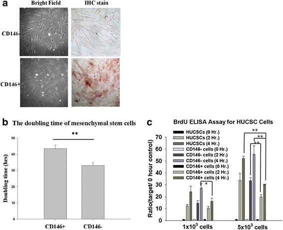
Phenotypes of expanded CD146+ and CD146– MSCs, and their proliferative properties. The phenotypes and anti-CD146 immunohistochemical staining of CD146+ and CD146– cells (magnification, 100×). a–c The proliferation rates of CD146+ and CD146– cells were detected by their doubling time and analyzed by BrdU ELISA. Data are expressed as mean ± SEM from five independent experiments. *P <0.05, **P <0.01. BrdU 5-bromo-2′-deoxyuridine, ELISA enzyme-linked immunosorbent assay, Hr hours, IHC immunohistochemistry