Fig. 3.

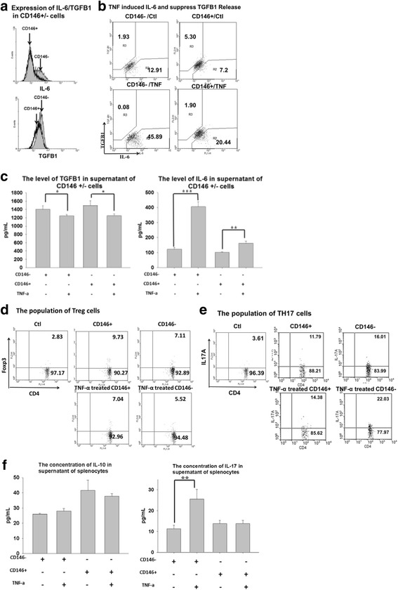
Immunomodulation of CD146+ and CD146– cells in vitro. IL-6 and TGF-β1 expression on CD146+ and CD146– cells was analyzed by flow cytometry and ELISA. MSCs were treated with or without 10 ng/ml TNFα for 3 days, and the concentrations of IL-6 and TGF-β1 were measured intracellularly and in the supernatants a–c. CD4+ Foxp3+ cells and CD4+ IL-17A+ cells were cocultured with TNFα-pretreated CD146+ cells or CD146– cells for 2 days. The T cells were analyzed by flow cytometry (d Treg cells; e Th17 cells) and ELISA (f left IL-10, right IL-17). Data are expressed as mean ± SEM from five independent experiments. *P <0.05, **P <0.01, ***P ≤0.001. Ctrl control, IL interleukin, TGF-β transforming growth factor beta, TH17 T-helper type 17, TNFα tumor necrosis factor alpha, Treg regulatory T
