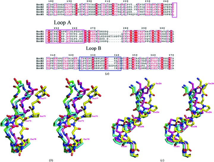
Figure 3.
(a) Sequence alignment of M. tuberculosis ESX-1–ESX-5 EccB homologues. Alignment of the sequences of EccB homologues from the ESX-1–ESX-5 secretion systems. Loops A (residues 243–264) and B (residues 324–341) are boxed in pink and blue. (b, c) Structure superimposition of loops A (b) and B (c) in the four states. Main-chain atoms and proline residues are shown as sticks. States I, II, III and IV are shown in green, blue, pink and yellow, respectively. N and C atoms are shown in blue and red, respectively.

