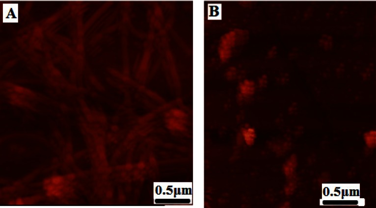
Figure 5.
AFM images of HEWL aggregates deposited on mica following inhibition by CE. AFM samples were taken when ThT fluorescence indicated that control samples containing HEWL alone had reached the equilibration phase. Control samples showed long and mature fibrils (A). HEWL in presence of 1 mg/ml CE after 48 hour incubation at 57 °C revealed the presence of small oligomers and disappearance of fibrils (B).

