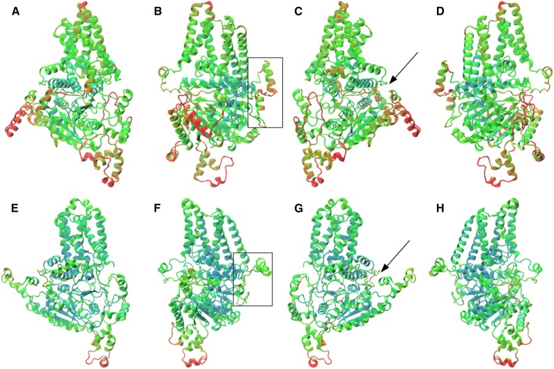
Figure 8.
Homology model (a–d) and final MD structure (e–h) of the barley HvCslF6 protein, which is believed to mediate (1,3;1,4)-β-glucan synthesis. The structures are shown progressively rotated 90° from left to right and colored by the root mean square fluctuation (RMSF) of each residue (blue = low fluctuation, red = high fluctuation). The homology model is colored by RMSF calculated over the entire 50-ns simulation, while the MD structure is colored by the final 10 ns only. The CslF6-specific insert of approximately 55 amino acids is highlighted in the boxes in b and f. The position of the TED/QxxRW motif of the active site is indicated with black arrows in c and g.

