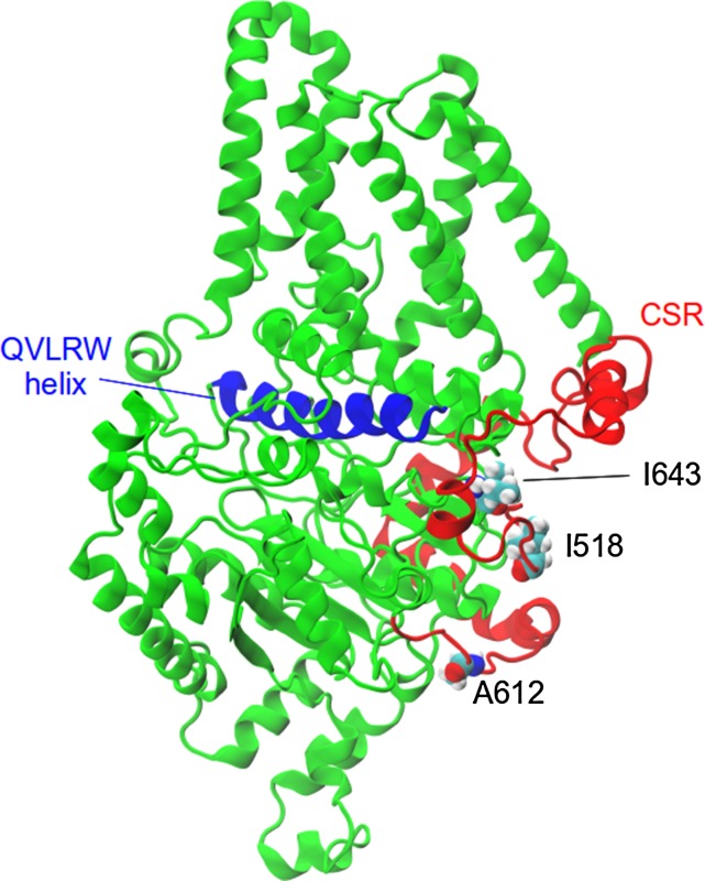
Figure 9.
Model of the barley CslF6 enzyme, showing the positions of the residues under selection (in a van der Waals representation). The CSR is colored red, while the helix that contains the QxxRW motif and sits above the active site is colored blue.

