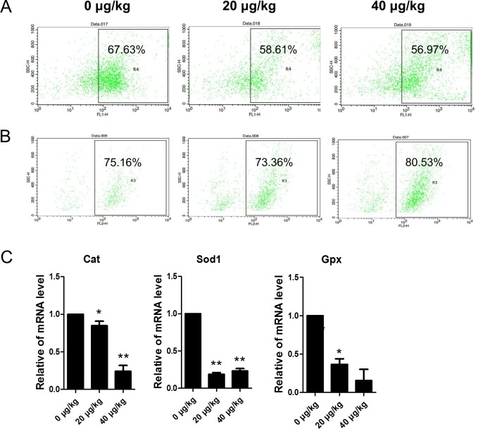
Fig 7. Effects of DEHP exposure on oxidative stress (DCF and DHE) and apoptosis in ovarian somatic cells.
(A-B) Flow cytometer analysis of the oxidative stress in (DCF and DHE) ovarian somatic cells in vivo. (C) The change of mRNA levels of related oxidative stress genes Cat, Sod1 and Gpx in the control and DEHP-treatment groups in in vivo experiments, respectively. Compared to the control group, relative fold changes were presented as mean ± SD. All experiments were repeated at least three times independently. (* P < 0.05; ** P < 0.01).

