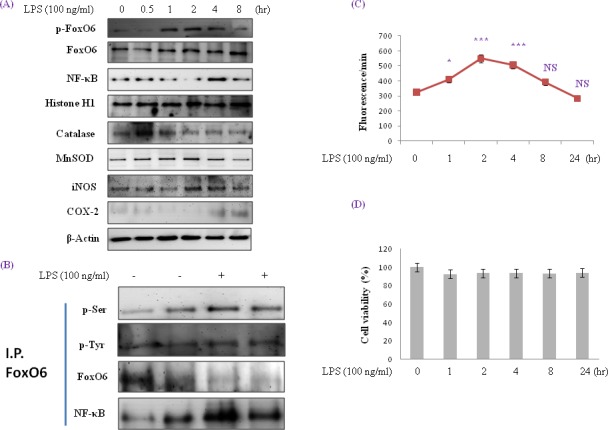
Figure 1. Enhancement of FoxO6 phosphorylation and NF-κB protein levels in LPS-treated HepG2 cells.

HepG2 cells were treated with 100 ng/ml LPS for various times. Samples loaded on sample gel were probed with β-actin and histone H1. A. Nuclear levels of FoxO6 and NF-κB and cytoplasmic levels of catalase, MnSOD, iNOS, and COX-2 were noticeably diminished after LPS treatment for 0.5 to 8 hr. B. Immunoprecipitated FoxO6 was found to be physically associated with NF-κB by Western blotting. C. Quantitative analysis was performed by measuring DCFDA fluorescence after treating plate with vehicle or 100 ng/ml LPS for 1 to 24h. Results were obtained using one-factor ANOVA: ***p < 0.001 vs. LPS untreated cells. D. Cells were treated with 100 ng/ml LPS for 24hr. The MTT assay used is described in Materials and Methods.
