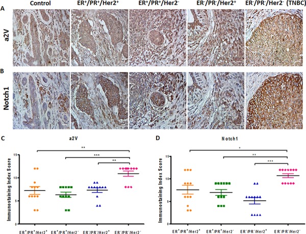
Figure 3. a2V and Notch1 are activated in human breast tumors.

Tissue microarray containing human breast tumors and normal breast tissues (control) were used to immunolocalize A. a2V and B. Notch1. Tumors were grouped by receptor-defined subtype. 12 sections per subtype were analyzed. Brown staining - DAB, counterstain - hematoxylin. Original magnification: 400X. Corresponding scatter dot plots show immunostaining index score ((ISIS) = Stained area score (SAS) × Immunostaining intensity score (IIS)) for C. a2V and D. Notch1. Data represent mean ± standard error, n = 12. *P ≤ 0.05, **P ≤ 0.01, ***P ≤ 0.001 between a pair of subtypes. DAB: 3,3′ Diaminobenzidine.
