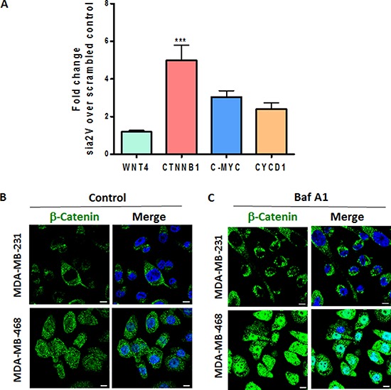
Figure 6. a2V-ATPase inhibition enhances Wnt signaling in TNBC.

A. MDA-MB-231 cells were transfected with scrambled control or a2V siRNA and harvested after 48 hrs of transfection. Fold change in mRNA expression levels of Wnt signaling genes WNT4, β-catenin (CTNNB1), C-MYC and Cyclin D1 (CYCD1) was assessed by qRT PCR. Prior to fold--change calculation, the values were normalized to signal generated from endogenous control 18srRNA. Data represent mean ± standard error, n = 4. *P ≤ 0.05, **P ≤ 0.01 compared to control siRNA. (B and C) TNBC cells were grown on chamber slides and treated with B. vehicle control or C. 0.1 μM Baf A1 for 4 hours. Cells were fixed, permeabilized and processed for immunofluorescence microscopy. Localization of β-catenin (green) is shown. Nucleus was stained with DAPI (blue). Scale bars: 10 μm.
