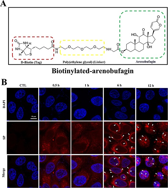
Figure 7. The cellular localization of arenobufagin in live cells.

A. The chemical structure of biotinylated arenobufagin. B. HepG2 cells were incubated with biotinylated arenobufagin for various times and then probed with SP. Nuclear DNA was stained with DAPI. Arrows indicated the granules of accumulation of biotinylated-arenobufagin. Original magnification: 630×; Scale bar: 10 μm.
