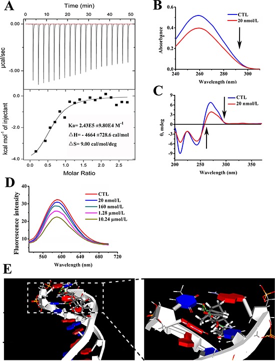
Figure 8. Arenobufagin directly binds with DNA via intercalation.

A. Arenobufagin binding to DNA was measured by ITC. A total of 30 μmol/L of DNA was titrated with 0.4 mmol/L of arenobufagin. The resulting thermograms were analyzed based on the one set of binding sites model using Microcal Origin 7.0 (Microcal. Inc.). B. The effect of arenobufagin on the UV absorption spectrum of DNA. 1 mmol/L DNA solution was mixed with 20 nmol/L arenobufagin. After the solution was mixed and equilibrated for approximately 5 min, the absorption spectra were measured at wavelengths ranging from 200 nm to 400 nm. C. The effect of arenobufagin on the CD spectra of DNA. The CD spectra of DNA (1 mmol/L) in 50 mmol/L Tris-HCl (pH = 8.0) with 20 nmol/L of arenobufagin. Each spectrum was analyzed from 200 nm to 370 nm at 25°C with a 10 mm path length cell. D. Fluorescence titration of EB-DNA complex with arenobufagin. EB-DNA complex was excited at 524 nm, and emission spectra was recorded from 530 to 700 nm at 25°C. E. The docked conformations suggested the intercalation between arenobufagin and d(CCGGCGGT)2. The green dotted lines represent the hydrogen bonds formed between arenobufagin and the DNA duplex.
