
Figure 5. Structure of the Zn-RING-Zn domain of LNX2.
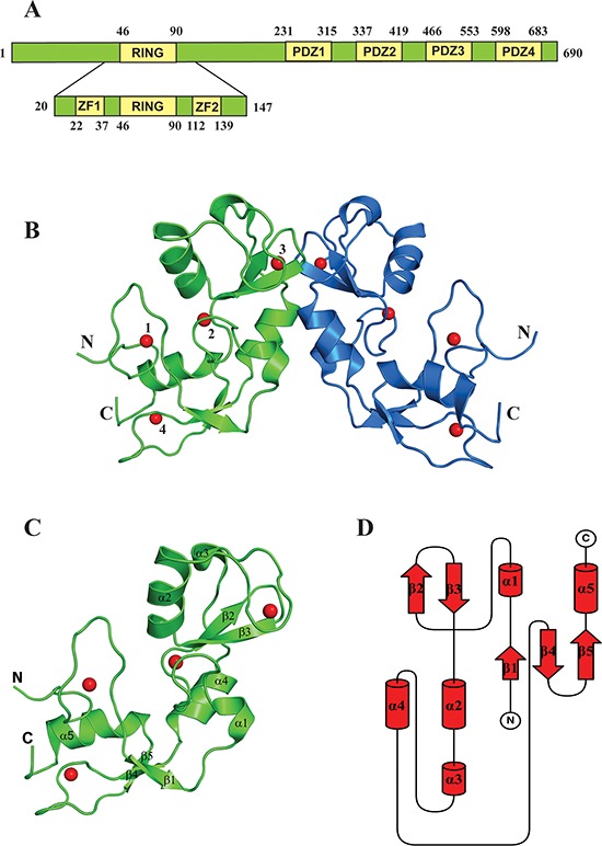
A. Schematic representation showing the domain architecture of the full-length LNX2 (above) and the Zn-RING-Zn domain of LNX2 (below). ZF = Zinc finger domain. B. The crystal structure of the Zn-RING-Zn domain of LNX2 (aa 20-147). Each monomer contains four zinc coordination sites. Sites 2 and 3 are located in the RING domain. Sites 1 and 4 are coordinated by two Zinc finger motifs, respectively. C. Structure of the monomeric Zn-RING-Zn domain along with four coordination Zinc ions shown as red spheres. α1 and α3 are single turn helix. D. Topology diagram of the Zn-RING-Zn domain of LNX2, as described in (C).