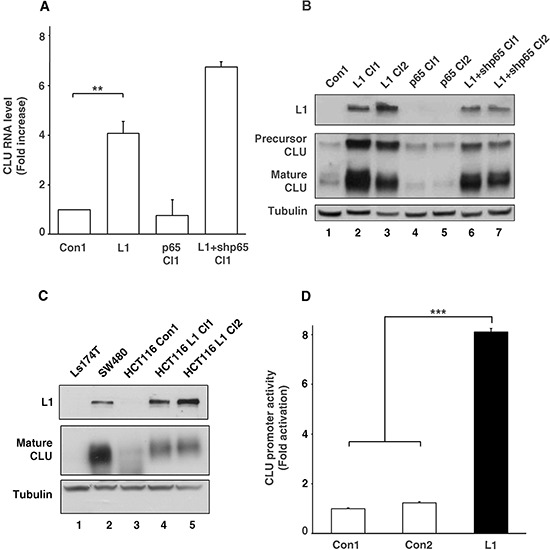
Figure 1. Clusterin (CLU) expression is elevated by L1 in CRC cells by CLU promoter activation independently of the NF-κB pathway.

A. RNA was extracted from individual cell clones isolated from stably transfected Ls174T cells with L1, control plasmid (Con1), L1 together with shRNA against p65 (L1+shp65 Cl1), and p65 alone (p65 Cl1). Quantitative RT-PCR was conducted using primers for CLU and GAPDH as control. B. Western blot analysis for L1, CLU and Tubulin as loading control, of the cell clones shown in A. Two cell clones of each type were used except for the control. C. Western blot analysis of the conditioned medium (CM) from Ls174T, SW480, HCT116 cells and HCT116 cell clones stably transfected with L1 (lanes 4 and 5). D. The CLU gene promoter reporter plasmid was co-transfected together with pSV β-galactosidase (as control vector for transfection efficiency normalization) into Ls174T CRC cells stably transfected with L1 and into two controls: a non-transfected Ls174T control and a Ls174T clone transfected with pcDNA3 (Con1 and Con2, respectively). Fold CLU promoter activation was determined after dividing luciferase activity by the values obtained with the empty reporter plasmid (pA3 vector). **p < 0.01, ***p < 0.001. Error bars: ±S.D.
