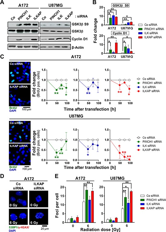
Figure 4. Integrin-associated proteins affected cell cycle and DNA double strand repair.

A. Western blot and B. densitometric analysis of p53-wildtype A172 and U87MG cells 48 h after PINCH1, ILK or ILKAP knockdown. Non-specific siRNA served as the control. C. Percentage of BrdU positivity as a marker for S-phase cells at different time points after siRNA transfection. Representative images are shown. D. Immunofluorescence staining of 53BP1 (green) and γH2AX (red) and E. quantitative analysis of foci numbers in irradiated A172 and U87MG cells after PINCH1, ILK or ILKAP depletion. Nuclei were stained with DAPI (blue). Data are mean ± SD (n = 3; t-test; *P < 0.05, **P < 0.01).
