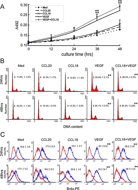
Figure 3. CCL18 promoted HUVEC angiogenesis without enhancing proliferation.

A. MTT assays for HUVECs cultured in media with or without rCCL18 (20 ng/mL), rCCL20 (20 ng/mL), rVEGF (10 ng/mL), or combined rCCL18 (20 ng/mL) and rVEGF (10 ng/mL) for 12, 24, 36, or 48 h. All values are means ± SEMs from 4 independent experiments. **p < 0.01 versus the media-only group. B. FACS analysis of cell cycle distribution in HUVECs cultured with or without rCCL18 (20 ng/mL), rCCL20 (20 ng/mL), rVEGF (10 ng/mL), or combined rCCL18 (20 ng/mL) and rVEGF (10 ng/mL) for 24 or 48 h. The histograms represent data from 4 independent experiments. All values are means ± SEMs from 4 independent experiments. **p < 0.01 versus the media-only group. C. BrdU-incorporation assays performed by flow cytometry in HUVECs treated as described in (B), using a PE-conjugated anti-BrdU antibody. The histograms represent 4 independent experiments. Means ± SEMs of fluorescence intensities for BrdU-treated HUVECs (blue) versus untreated cells (red) are indicated in each panel. **p < 0.01 versus the media-only group.
