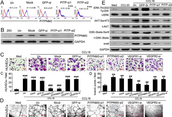
Figure 5. CCL18 enhanced HUVEC migration and tube formation via PITPNM3.

A. Flow-cytometric analysis of PITPNM3 (PITP) expression (blue) relative to an IgG isotype control (red) in HUVECs that were untreated (Un), mock-transfected, or transfected with either of 2 PITPNM3 siRNAs or a GFP-siRNA. Mean fluorescence intensities ± SEMs for PITPNM3 immunostaining are indicated for 3 independent experiments. ***p < 0.001 versus untreated cells. B. Representative western blot results for PITPNM3 (PITP) expression in HEK293 cells and HUVECs transfected as described in (A). GAPDH was used as a loading control. C–D. Migration assays (C) and Matrigel tube-formation assays (D) in CCL18-treated HUVECs pretransfected with GFP, PITPNM3, VEGFR1, or VEGFR2 siRNAs. Scale bar, 50 μm. Bars correspond to means ± SEMs from 3 independent experiments. **p < 0.01 and ***p < 0.001 versus the media-only group; ##p < 0.01 and ###p < 0.001 versus the untransfected control (Un). E. Representative western blot results showing the phosphorylated and total levels of the ERK, AKT, GSK-3β, and Snail proteins in HUVECs treated with medium alone, rCCL20 (20 ng/mL), or rCCL18 (20 ng/mL), with or without transfection of either of 2 PITPNM3 (PITP) siRNAs or a GFP-siRNA. GAPDH was detected as a loading control.
