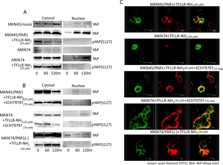
Figure 4. PAR1 promotes YAP dephosphorylation.
The left line panels are immunoblotting of cytoplasmic lysates. The right panels are immunoblotting of nucleus lysates. A. Both MKN45/PAR1 and MKN74 cells were treated with TFLLR-NH2 for indicated periods of time. Cytoplasmic and nuclear cell lysates were separated. And these cells lysates were subjected to immunoblotting with the YAP1 and pYAP1 (S127). B. Both MKN45/PAR1 and MKN74 cells were treated with TFLLR-NH2 and SCH79797 for indicated times. MKN74/PAR1(−) cells were treated with TFLLR-NH2 for indicated times. These cell lysates were also separated cytoplasmic and nucleus and subjected to immunoblotting with the YAP1 and pYAP1 (S127). C. Immunofluorescence staining with anti-actin (green) and anti-YAP1 (red) antibodies; the right panels show the overlay of the green and red staining.

