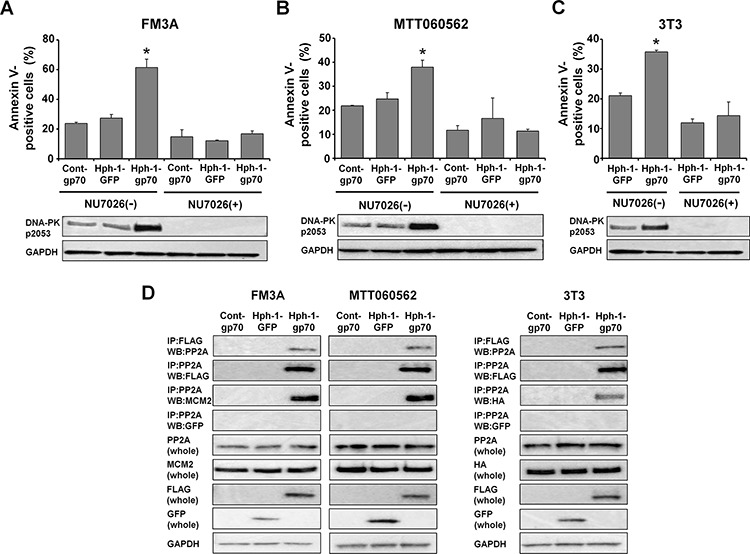
Figure 5. The Hph-1-gp70-MCM2 complex binds to PP2A and causes hyperphosphorylation of DNA-PK.

A. FM3A cells and B. MTT060562 cells were pre-incubated with or without 10 μM NU7026, a DNA-PK-inhibitor, for 2 h and treated with 1 μM cont-gp70, Hph-1-GFP, or Hph-1-gp70 and 500 nM doxorubicin for 24 h. The apoptotic cell ratios were determined 24 h later with annexin V-staining (top). *P < 0.01 by two-tailed Student's t-test. DNA-PK-pS2053 levels were analyzed by western blotting (bottom). C. HA-Mcm2-expressing 3T3 cells were pre-incubated with or without 10 μM NU7026 for 2 h and treated with 1 μM Hph-1-GFP or Hph-1-gp70 and 500 nM doxorubicin for 24 h. The apoptotic cell ratios were determined 24 h later with annexin V-staining (top). *P < 0.01 by two-tailed Student's t-test. DNA-PK-pS2053 levels were analyzed by western blotting (bottom). D. FM3A (left), MTT060562 (middle), and HA-Mcm2-transfected 3T3 cells (right) were treated with 1 μM cont-gp70, Hph-1-GFP, or Hph-1-gp70 for 2 h. Cell lysates were subjected to pull-down assays to assess the binding of PP2A to gp70 and PP2A to MCM2.
