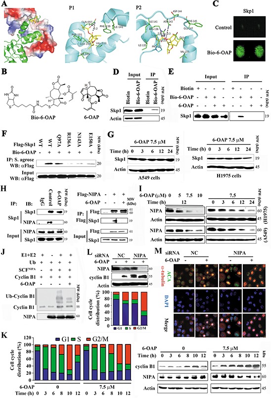
Figure 2. 6-OAP directly binds Skp1 and interferes with SCFNIPA.

A. Two potential binding pockets (P1 and P2) of Skp1 for 6-OAP, revealed by docking 6-OAP to Skp1 (PDB code: 2AST). In the left panel, Skp2 (shown as cartoon) interacts with Skp1 (shown as surface) via P1 and P2, and only Skp1 is used during the docking. In the middle and right panels, 6-OAP (shown as sticks) is predicted to interact with Skp1 (shown as cartoon) via P1 and P2, respectively. B. Chemical structure of 6-OAP and Bio-6-OAP. C. Images of Bio-6-OAP-Skp1 interaction on the slides. D. H1975 cells were treated with Biotin or Bio-6-OAP at 50 μM for 6 h, lysed, and the cell lysates were subjected to immunoprecipitation using streptavidin agarose and Western blot using indicated antibodies. E. H1975 cells were treated with Bio-6-OAP (50 μM) in the presence or absence of 6-OAP (100 μM) for 6 h, lysed, and the cell lysates were subjected to immunoprecipitation and Western blot. F. 293T cells were transfected with wild type (WT) or mutant Skp1 for 48 h, lysed, the lysates were subjected to immunoprecipitation using streptavidin (S.) agarose and Western blot using indicated antibodies. G. The cells were treated with 6-OAP, lysed, and subjected to Western blot. H. H1975 cells were treated with or without 6-OAP for 3 h, lysed, and immunoprecipitation and Western blot assays were performed (left panel). 293T cells were transfected with pcDNA3.1-flag-NIPA, treated with or without 6-OAP, and lysed for immunoprecipitation and Western blot (right panel). I. Cells were treated with 6-OAP, lysed, and Western blot was performed. J. An in vitro ubiquitination assay using SCFNIPA, Cyclin B1, and 6-OAP. K. A549 cells were synchronized to G1/S boundary and released, and treated with or without 6-OAP. Cell cycle distribution was determined (left), and the expression of NIPA and Cyclin B1 was analyzed by Western blot (right). L. A549 cells transfected with control or NIPA specific siRNA were treated with 6-OAP for 12 h, harvested for Western blot (upper) or flow cytometry analysis (lower). M. A549 cells transfected with NIPA-specific siRNA were treated with or without 6-OAP (7.5 μM) for 12 h, and analyzed by immunofluorescence labeling with anti-centromere sera, anti-α-tubulin antibody, and DAPI. Size bar, 5 μm.
