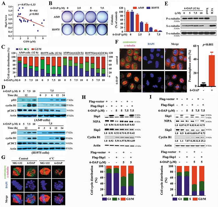
Figure 4. 6-OAP induces prometaphase arrest in lung cancer cells.

A. The GI50s of 6-OAP in cells is associated with the relative Skp1 expression. B. Effects of 6-OAP on clonogeinc activity of lung cancer cells. C. The synchronized or asynchronous (asyn) to G1/S boundary cells were treated with 6-OAP at indicated concentrations for 12 h. Cell cycle distribution was determined by propidium iodide (PI) staining and flow cytometry analysis. D. The cells were treated with 6-OAP, lyzed, and Western blot was performed using antibodies indicated. E. The cells were treated with 6-OAP, taxol (50 nM) or Vincristine (VCR; 50 nM) for 12 h. The polymerized (P) and soluble (S) tubulin fractions were prepared and subjected to Western blot using anti-α-tubulin antibody. T-α-tubulin, total-α-tubulin. F. A549 cells were treated with 7.5 μM 6-OAP for 12 h, and assayed by immunofluorescence labeling with anti-centromere sera (green), anti-α-tubulin antibody to visualize microtubules (red), and DAPI to counter stained DNA (blue). Size bar, 5 μm. G. A549 cells were treated with 7.5 μM 6-OAP for 12 h or 10 μM MG-132 for 3 h, and incubated in ice-cold media for 10 min and stained with anti-centromere sera, anti-α-tubulin antibody, and DAPI. H, I. The A549 (H) and 293T (I) cells were transfected with Skp1, synchronized at G1/S boundary site by thymidine treatment, and treated with 6-OAP for 12 hours. The cells were analyzed by flow cytometry to evaluate the cell cycle distribution, or lysed for Western blot analysis. Numbers under the NIPA, Skp2, and Cyclin B1 bands are the relative expression values to Actin determined by densitometry analysis. *p = 0.04.
