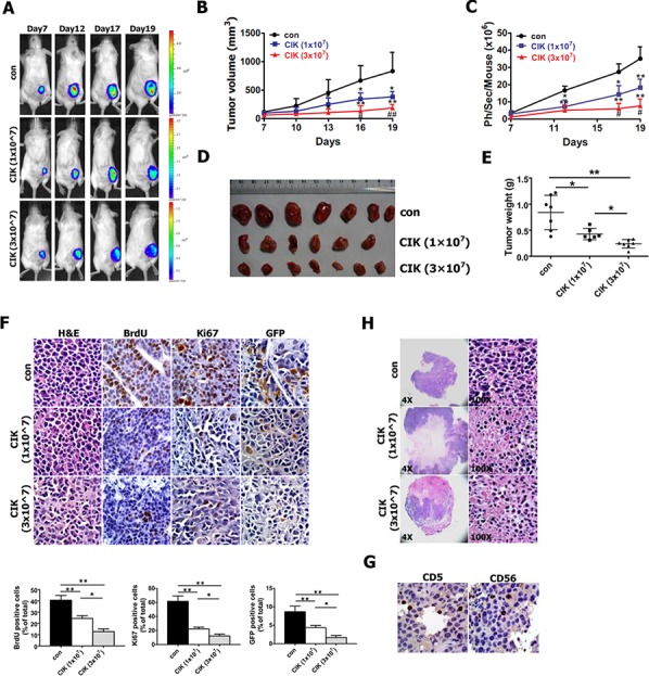
Figure 7. In vivo activity of CIK cells against CNE2 cells harboring GFP-T2A-Luc in NOD/SCID mice.

As mentioned in Materials and methods section, NOD/SCID mice were subcutaneously implanted with 1 × 10⁶ CNE2 cells harboring PNanog-GFP-T2A-Luc transgene. 7 days after tumor cell implantation, 1 × 10⁷ and 3 × 10⁷ CIK cells were infused by tail vein injection into recipient mice once every two days. A. Series of in vivo bioluminescence images (taken at the indicated times) of 3 representative NOD/SCID mice from CIK- (1 × 10⁷), CIK- (3 × 10⁷) and vehicle-treated groups before and after CIK cell treatment. B. Growth curve of tumor volumes. C. Quantification analysis of bioluminescence signal of tumor-bearing mice treated with CIK cells (1 × 10⁷ and 3 × 10⁷) or with vehicle control. D. Representative picture of tumors formed. E. Tumors were weighted. F. BrdU, Ki67 and GFP-stained sections of transplanted tumors formed by CNE2 cells at 19 days after subcutaneous transplantation. The percentages of BrdU-, Ki67- or GFP-positive cancer cells were calculated by the total number of BrdU-, Ki67- or GFP-positive cells over total number of cancer cells. G. Infiltration of CIK cells at tumor sites was shown by immunohistochemistry (Ab anti-CD5 and Ab anti-CD56) at the end of experiment. H. A significant increase in transplanted tumor tissue necrosis and apoptosis was observed in CIK-treated mice compared with controls at the end of the experiment.
