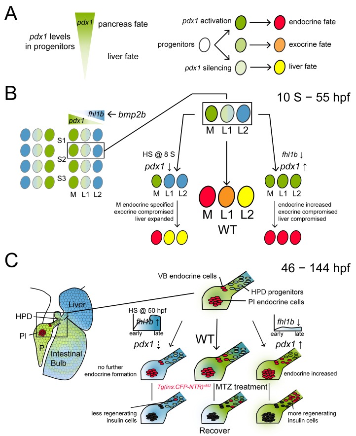
Fig 7. Fhl1b is essential for regulating the cell fate choice of liver versus pancreas and for β-cell regeneration.
Schematic model for the role of Fhl1b in lineage specification and in β-cell regeneration. The expression of fhl1b and pdx1 is color-coded as blue and green, respectively. (A) Endodermal progenitors experience different levels of pdx1 regulating their fates as pancreatic endocrine (high levels of pdx1), pancreatic exocrine (low levels of pdx1), or liver (pdx1 silencing) cells. (B) From the 12-somite stage onwards, single endodermal cells in the lateral 2 position (L2) between somites 1 and 3 in the endodermal sheet give rise not only to pancreatic exocrine cells and intestinal cells, but also to liver cells, functioning as bipotential hepatopancreatic progenitors. bmp2b, which is expressed in the lateral plate mesoderm, induces fhl1b expression in the prospective liver anlage. This may form a reciprocal gradient of fhl1b-pdx1. Decreased Fhl1b function leads to an increase in levels of pdx1 expression in lateral 1 and 2 cells, causing a hepatic and pancreatic exocrine to a pancreatic endocrine fate switch. Conversely, augmentation of Fhl1b activity at the initial time point of pdx1 expression in the pancreatic exocrine and intestinal progenitors (HS @ 8 S) causes a decrease in levels of pdx1 expression in pancreatic exocrine and intestinal progenitor cells, leading them to become liver cells. The lineage of the most medial cells, which express high levels of pdx1 and subsequently give rise to pancreatic endocrine cells, is specified primarily during the gastrulation stage. (C) At later embryonic/larval stages, the HPD system comprises a progenitor cell population that can differentiate into pancreatic endocrine cells and liver cells. At these stages, fhl1b (color-coded as blue) shows a reciprocal expression pattern with pdx1 (color-coded as green). Liver cells, which never express pdx1, express high levels of fhl1b, while the HPD system expresses low levels of fhl1b. The distal intestine also expresses fhl1b, whereas most pancreatic cells except for a few cells in the principal islet do not express fhl1b. In normal development, the ventral bud-derived β-cell formation initiates between 40–46 hpf (VB endocrine cells). Overexpression of fhl1b inhibited further induction of pancreatic endocrine cells and β-cell regeneration by potentially inhibiting pdx1 expression in the HPD system, whereas suppression of fhl1b increased pdx1 and neurod expression in the HPD system, augmenting pancreatic endocrine cell formation and subsequent β-cell regeneration. Abbreviations: S1, somite 1; S2, somite 2; S3, somite 3; M, medial; L1, lateral 1; L2, lateral 2; HPD, hepatopancreatic duct; PI, principal islet; P, pancreas; VB, ventral bud; WT, wild-type; MTZ, metronidazole.

