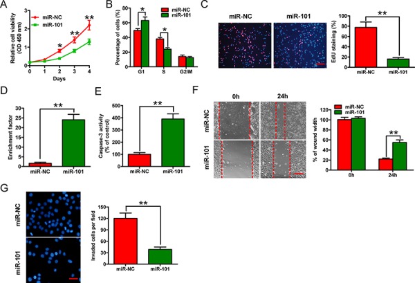
Figure 2. Effects of miR-101 on in vitro proliferation, apoptosis, migration, and invasion of BrC cells.

The 4T1-luc2-M cell line was transfected with 100 nM miR-101 mimic or the negative control miR-NC mimic. A. CCK-8 assay of cells transfected with miR-101 or miR-NC for the indicated number of days. B. Fluorescence-activated cell sorting assay of cells transfected with miR-101 or miR-NC, showing the effects on cell cycle progression. C. EdU incorporation assay of cells transfected with miR-101 or miR-NC. Scale bar = 20 μm. D, E. Nucleosomal fragmentation (D) and caspase-3 activity (E) assays of cells transfected with miR-101 or miR-NC. F, G. Wound-healing (F) and transwell (G) assays of cells transfected with miR-101 or miR-NC showing the effects on migration and invasion of 4T1-luc2-M cells. Scale bars = 10 μm (migration) or 20 μm (invasion). (C, F, G) Representative images are shown. (A–G) Data are represented as the mean ± SD of three replicates. *P < 0.05 and **P < 0.01.
