
Figure 2. B5 induced apoptosis and cell cycle arrest in CaSki and SiHa cells.
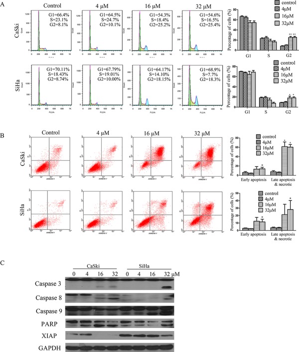
A. Effects of B5 on cell cycle distribution. Cells were treated with 0, 4, 16, and 32 μM B5 for 48 h, fixed in 70% ethanol, stained with PI, and cell cycle distribution was assessed by flow cytometry. The percentage of cells in each cell cycle phase is indicated as the mean ± SD of three independent experiments. B. Induction of apoptosis by B5 in CaSki and SiHa cells. Cells were treated as above and analyzed by flow cytometry after Annexin-V-FITC/PI staining; data are presented as the mean ± SD of three independent experiments. *P < 0.5 and **P < 0.01. C. Western blotting analysis of the expression of the following: pro- and cleaved-caspases 3, 8, and 9; PARP (86kDa); and XIAP. Data are representatives of three independent experiments.