
Figure 5. Effects of B5 on the Trx system.
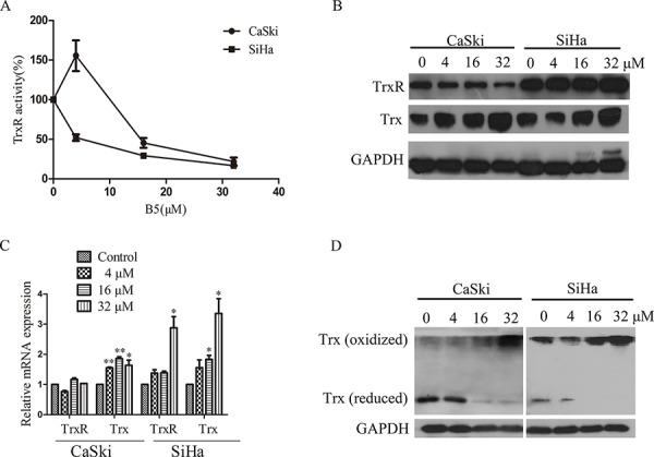
Cells were treated with 0, 4, 16 and 32 μM B5 for 48 h. A. TrxR activity in cell lysates was determined by SC-TrxR assay. Data were presented as the percentage of control. B. Trx and TrxR protein levels were analyzed by SDS-PAGE and western blotting. C. The mRNA levels of Trx and TrxR were measured by RT-PCR; GAPDH was used as an internal control. D. Trx redox status was determined by non-reduced SDS-PAGE and western blotting analysis; GAPDH was used as the internal control. All data are representatives of three independent experiments.