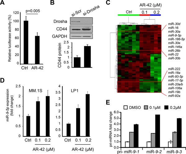
Figure 3. AR-42 upregulates expression of miR-9-5p.

A. Luciferase assay in MM.1S cells transiently transfected with pGL3-CD44 3′UTR construct and treated for 24 hrs with 0.2 μM AR-42, or vehicle control (Ctrl) showing inhibitory response to AR-42 via 3′UTR element. Each measurement was done in triplicate. B. MM.1S cells were treated with RNA silencing for Drosha (si-Drosha) or unrelated sequence (si-Scr). Forty eight hours later, cells were lysed and analyzed by western blot using anti-Drosha and anti-CD44 antibodies. GAPDH was used for normalization. Signals were quantified using ImageJ and plotted below. C. Dendrogram of the unsupervised hierarchical clustering analysis of global miRNA expression in MM.1S cells treated with designated concentrations of AR-42, or vehicle control (Ctrl), using NanoString technology. Selected most up-regulated (upper) and down-regulated (lower) miRNAs are indicated. D. miR-9-5p expression in MM.1S (left) and LP1 (right) cells treated with AR-42 at 0.1 and 0.2 μM, or vehicle control (Ctrl) was determined by qRT-PCR. Results are expressed as fold change compared to the DMSO (Ctrl). E. The effect of 24-hr treatment of MM.1S cells with AR-42 (at indicated concentrations) on expression of pri-miR-9-1, pri-miR-9-2 and pri-miR-9-3 was determined by qRT-PCR.
